There are two types of stonefish - estuarine and reef. They are brown to sandstone in colour, which allows them to blend in with their surroundings. Envenomation occurs when the fish is stood on. Stonefish have 13 dorsal spines (each with an attached venom gland) that are used defensively, not to catch food. When the stonefish is stepped on, the spines are pushed down and act like an injector, forcing venom into the wound. After envenomation, the puncture mark has a bluish tinge around it. The severe, local pain extends to the rest of the limb, causing severe pain and paraesthesiae (a tingling or burning sensation of the skin). The victim experiences low blood pressure, pale skin, nausea and dizziness. Other fish that are venomous include the bullroot, lungfihs, scorpion fish, and lionfish (Fenner, 2004).

A stonefish (Image 1).

A reef stonefish camouflaged to look like coral (Image 2).
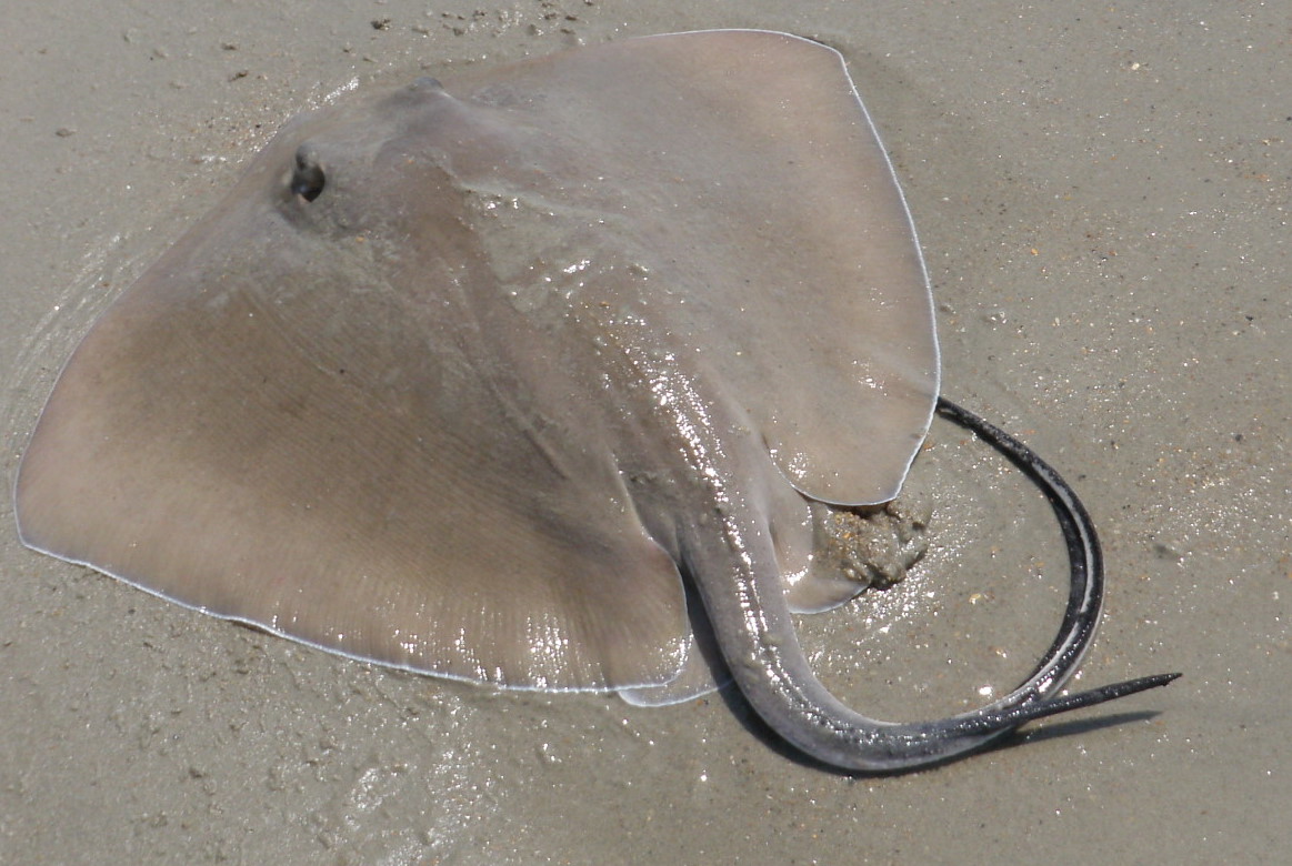
Stingrays are large, flat fish with flaps that enable them to swim gracefully through water. They have a tail with at least one barb on it, but they can have up to seven barbs present. The barbs face backwards and are covered in a friable sheath of tissue. They are often accidentally stood on, as they bury themselves in the sand to rest. When they are stepped on, the stingray's tail whips forward and the barb/barbs easily penetrate the skin of the victim. The pain is instant and severe. Jagged lacerations can occur, and may cause heavy bleeding. The barbs can often break off, become embedded in the tissue or be cleanly withdrawn after the predator was attacked. After the barb penetrates the tissue, the sheath that covers it is torn off, leaving a trail of venom and debris. Envenomation causes localised myolsis (the breakdown of muscle tissue) and inflammation, with local or extended tissue necrosis. If the abdomen or chest of the predator was punctured, there is an increase in morbidity and mortality (Fenner, 2004).

A stingray (Image 3).
Sea urchins are covered in hundreds of sharp spikes. They live on rocks close to or in shallow waters. When they are stood on, the spikes break off into the victim's foot. The sea urchin's venom is not very toxin, but causes an unpleasant pain (Fenner, 2004).

A sea urchin (Image 4).
Cone shells are brightly coloured, triangular-shaped shells, with
a longitudinal fold or split running the length of the shell. They have a proboscis (a small, hollow, flexible tube) that can
emerge from anywhere along the slit. At the base of the proboscis is an area
containing a number of barbs, called radicular teeth. These barbs are bathed in a potent venom and are fired at prey and predators. Envenomation causes a localised, sharp pain. There is very little reaction from the body. Some flushing of the skin may be present, and sometimes a rash can form. The worst reactions occur when someone is stung by a fish eating cone, as the venom is more intense. The venom rapidly causes numbness and local swelling,
nausea, incoordination, muscular weakness and difficulty breathing due to
weakness of the diaphragm and intercostal muscles. Envenomation may lead to respiratory paralysis and death from
asphyxia (Fenner, 2004).


Sea snakes are common in all oceans (except the Atlantic), but are most common in tropical and sub-tropical Australia. They are similar in appearance to land snakes, except they have a flattened, paddle-like tail to help them swim. They do not have gills, and so must surface to breathe air. They can be divided into two groups - those that have large mouths but rarely bite or envenomate, and those that have small mouths and very potent venom, but are unable to take a large enough bite to envenomate most animals. However, if something is even scratched and envenomated by the extremely potent venom, death is almost certain to occur. Like land based snakes, sea snakes try to preserve their venom, and hence, most bites are dry (don't inject venom). The bite is relatively painless, but the venom causes drowsiness, nausea and vomiting, weakness, visual disturbances, breathing problems and muscles pains or stiffness (Fenner, 2004).
A cone shell with its proboscis extended (Image 5).
A cone shell striking a fish (Image 6).
Sea snakes are common in all oceans (except the Atlantic), but are most common in tropical and sub-tropical Australia. They are similar in appearance to land snakes, except they have a flattened, paddle-like tail to help them swim. They do not have gills, and so must surface to breathe air. They can be divided into two groups - those that have large mouths but rarely bite or envenomate, and those that have small mouths and very potent venom, but are unable to take a large enough bite to envenomate most animals. However, if something is even scratched and envenomated by the extremely potent venom, death is almost certain to occur. Like land based snakes, sea snakes try to preserve their venom, and hence, most bites are dry (don't inject venom). The bite is relatively painless, but the venom causes drowsiness, nausea and vomiting, weakness, visual disturbances, breathing problems and muscles pains or stiffness (Fenner, 2004).

A sea snake (Image 7).

A blue-ringed octopus (Image 8).

A diagram of an octopus's beak and mouth (Image 9).
References
Fenner, P. (2004). Venomous marine animals. South
Pacific Underwater Medicine Society Journal, 34 (4), 196-202.
Images
Image 1 - http://upload.wikimedia.org/wikipedia/commons/5/57/Stone_Fish_at_AQWA_SMC2006.jpg. Accessed on 31/3/14.
Image 2 - http://upload.wikimedia.org/wikipedia/commons/b/b8/Reef_Stonefish.jpg.
Image 3 - http://lenebaxter.files.wordpress.com/2012/04/stingraystinger.jpg. Accessed on 31/3/14.
Image 4 - http://www.gambassa.com/GambassaFiles/Images/images/JOHNpetrucci/urchin_V1.jpg. Accessed on 31/3/14.
Image 5 - http://grimwade.biochem.unimelb.edu.au/cone/Conus_textile-Paul_Livett.jpg.
Image 6 - http://www.rudyrucker.com/blog/images/sciamcone.jpg. Accessed on 31/3/14.
Image 7 - https://blogger.googleusercontent.com/img/b/R29vZ2xl/AVvXsEj4fl_k6nMjD2X509QMyaiHmNS7hqDeD1AED2OhXDa0NpJ97m_fUrSdGyU2bfNLfpFTt-6sk0pu11GQFoQ5s9t6z7yxJGkywDimT2w7amD3TeD4yrW6BNTmo7uwMY2vg79pN4NV2eZjihg/s640/ular+laut+2.jpg.
Image 8 - https://blogger.googleusercontent.com/img/b/R29vZ2xl/AVvXsEgZxjFbokMHogT8BUVozupgem6arvyQV1j3HmyES7brGSUVLvgxx4a6FZYX5EdlqEdI-VDaE4ca17y31SiQ5irGq9c759e-dgLXdwbjvcq7u1aE_D3jf_A7-1Xog9pbY47KDSewz50B06g/s1600/HLfemalesmoothdisplaymed.jpg.
Image 9 - https://blogger.googleusercontent.com/img/b/R29vZ2xl/AVvXsEjAwYCfCpy0MVKCjaCTsTpvDghjv53NEIYyfoSF-Oh1T5xaHdMiLAhaIL2QOUx1o0kzD08dknw-hJFMCGWv2Dx4A6AoG-azPhMfBYtrQhv1qctY_P97AbH3wkczHMv6QVs9WjZDTyX56FY/s400/octopus-2.jpg.


